Cardiac Cycle for FMGE: One Sequence, Not a List of Disconnected Facts
By Dr. Utsav Bhattacherjee, MBBS, MBA · 17 September 2026 · 11 min read
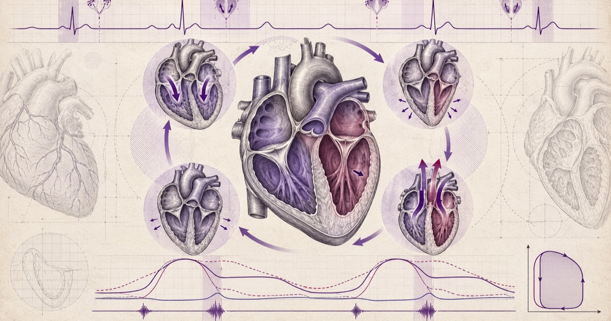
The cardiac cycle is one continuous sequence, but exam questions almost always ask about it in fragments — a specific pressure at a specific moment, a specific heart sound tied to a specific valve event. The fastest way through these questions is holding the whole cycle as one mental sequence, so any fragment can be placed back into its correct position rather than recalled as an isolated fact.
The Two Halves: Systole and Diastole
Systole is the period of ventricular contraction and ejection; diastole is the period of ventricular relaxation and filling. At a normal heart rate, diastole occupies a meaningfully larger share of the cycle than systole — which matters clinically, since a rising heart rate shortens diastole disproportionately, directly reducing the time available for ventricular filling and coronary perfusion, both of which happen predominantly during diastole.
Isovolumetric Contraction: Pressure Rises, Volume Doesn't
The cycle begins, functionally, with isovolumetric contraction: the mitral and aortic valves are both closed, the ventricle is contracting, and pressure rises sharply while volume stays completely constant, since no blood can enter or leave a sealed chamber. This phase ends the instant ventricular pressure exceeds aortic pressure, forcing the aortic valve open.
Ejection: Volume Falls as Blood Leaves
Once the aortic valve opens, ejection begins — ventricular volume falls as blood is pushed into the aorta, while pressure in both the ventricle and aorta rise together initially, then fall as ejection slows. Ejection ends when ventricular pressure drops below aortic pressure, at which point the aortic valve closes.
Isovolumetric Relaxation: The Mirror of Contraction
With both valves closed again, the ventricle relaxes and pressure falls sharply while volume, once again, stays constant. This phase ends when ventricular pressure drops below atrial pressure, opening the mitral valve and beginning ventricular filling.
Filling: Rapid, Then Slow, Then Atrial Kick
Filling itself has three sub-phases worth knowing: rapid filling immediately after the mitral valve opens (blood rushes in along the pressure gradient), a slower diastasis phase as the pressure gradient equalises, and finally atrial contraction — the atrial kick — which tops off ventricular filling just before the next systole begins. The atrial kick contributes a genuinely small fraction of filling in a healthy heart at rest, which is exactly why atrial fibrillation, which abolishes coordinated atrial contraction, is often well tolerated at rest but becomes clinically significant during exertion, when that extra filling contribution matters more.
The Full Sequence, Side by Side
| Phase | Valves | Volume | Pressure Trend |
|---|---|---|---|
| Isovolumetric contraction | Both closed | Constant | Rising sharply |
| Ejection | Aortic open, mitral closed | Falling | Rises then falls |
| Isovolumetric relaxation | Both closed | Constant | Falling sharply |
| Rapid filling | Mitral open, aortic closed | Rising fast | Low, falling slightly |
| Diastasis | Mitral open, aortic closed | Rising slowly | Low, stable |
| Atrial contraction | Mitral open, aortic closed | Rising (small amount) | Small rise |
Heart Sounds, Mapped to the Cycle
S3, when present, occurs during rapid ventricular filling — it’s a genuinely normal finding in children and young healthy adults, but in an older adult it points toward a volume-overloaded or failing ventricle, since a stiff or overloaded ventricle produces an audible vibration as blood rushes in rapidly. S4 occurs during atrial contraction, immediately before S1, and it results from blood being forced into a ventricle that is abnormally stiff — a non-compliant ventricle, as seen in longstanding hypertension or hypertrophic cardiomyopathy, produces this sound as the atrial kick meets resistance.
Murmur Timing Follows Directly From the Cycle
Once the cycle’s phases are clear, murmur timing stops being a separate memorization task. A murmur occurring between S1 and S2 is a systolic murmur, occurring during ejection — aortic stenosis and mitral regurgitation are the classic examples, since both involve abnormal flow during the ejection phase itself. A murmur occurring between S2 and the next S1 is a diastolic murmur, occurring during filling — aortic regurgitation and mitral stenosis are the classic examples, involving abnormal flow during the filling phase.
The Jugular Venous Pulse: A Second Window Onto the Same Cycle
The jugular venous pulse waveform is worth learning alongside the cardiac cycle specifically because it’s measuring the same underlying events from the right atrium’s perspective, giving a second, complementary view. The a wave reflects atrial contraction — the same event producing S4 and the atrial kick on the left side. The c wave reflects the tricuspid valve bulging slightly into the atrium during the start of ventricular contraction. The x descent reflects atrial relaxation and the downward pull of the contracting ventricle. The v wave reflects venous filling of the atrium against a closed tricuspid valve during ventricular systole. The y descent reflects the tricuspid valve opening and blood rushing into the ventricle.
A cannon a wave — an unusually large a wave — occurs when the atrium contracts against a closed tricuspid valve, classically seen in complete heart block, where atrial and ventricular contraction become dissociated and occasionally coincide. This is a genuinely useful bedside finding that directly reflects the same electrical and mechanical timing the cardiac cycle describes.
The Actual Numbers Worth Knowing
Normal left ventricular pressure swings from close to 0 mmHg at the end of diastole up to roughly 120 mmHg at peak systole — the same peak the aorta reaches once the aortic valve opens, since the two chambers are briefly one continuous pressure system during ejection. Aortic diastolic pressure stays elevated, typically around 80 mmHg, because the closed aortic valve and the elastic recoil of the aortic wall maintain pressure between beats — this diastolic pressure is what actually drives coronary blood flow, since the coronary arteries fill primarily when the aortic valve is shut and the myocardium itself is relaxed enough not to compress them.
Left atrial pressure stays low throughout, typically in the single digits to low teens in mmHg, which is exactly why even a small rise in left atrial pressure — as seen in mitral stenosis or left heart failure — is clinically significant enough to back up into the pulmonary circulation and produce pulmonary congestion. The right side of the heart runs at roughly a fifth of these pressures throughout the cycle, since the pulmonary circulation it serves is a low-resistance system compared to the systemic circulation the left heart serves.
This pressure asymmetry between the two sides of the heart is worth holding onto as a standing fact, not just a cardiac-cycle detail: it’s the reason right heart pathology and left heart pathology tend to present differently, and the reason a right-sided pressure that would be entirely normal on the left side already represents significant elevation on the right.
How the Cardiac Cycle Is Tested on FMGE
The most common question format gives you one point on the cycle — a pressure value, an open or closed valve, a named sound — and asks what is true at that same instant. These are answered by locating the moment rather than by recall, which is why the phase sequence is worth knowing as a sequence rather than as a list of definitions.
The second format is splitting of the second heart sound, and it is worth treating as its own topic because it follows directly from the cycle rather than from anything new. S2 is aortic closure (A2) followed by pulmonary closure (P2). Inspiration increases venous return to the right ventricle, lengthens right ventricular ejection and delays P2 — so a split that widens on inspiration and narrows on expiration is physiological. A split that stays wide and does not move with respiration at all is the classic finding in an atrial septal defect, where the shunt keeps right-sided filling constant across the respiratory cycle. A split where P2 comes FIRST and the gap narrows on inspiration is paradoxical splitting, and it points to something delaying aortic closure — left bundle branch block or severe aortic stenosis.
Recognising which of those three patterns a vignette describes is usually enough to reach the diagnosis without any further reasoning, which is what makes it high yield for the marks it carries. Practising the format against FMGE previous year questions is the fastest way to see how narrow the variation actually is. For the subject around it, how to prepare Physiology for FMGE covers how cardiovascular physiology is weighted, and the FMGE Physiology subject page gathers the related topics.
Putting the Whole Sequence Together
Reading a cardiac cycle question efficiently means anchoring on whichever single fact is given — a heart sound, a murmur timing, a JVP wave, or a named phase — and walking from that anchor to the surrounding phases, rather than trying to recall the entire cycle from the start every time. Each of these findings is a window onto the same underlying sequence, and knowing which valves are open, which are closed, and whether volume is changing at that exact moment is what actually answers most of these questions, not memorized association alone.
A vignette stating a specific measured pressure — say, a left ventricular pressure of 8 mmHg at a moment when the mitral valve is open — is really just describing one specific point on this same cycle, and translating that number back into which phase it belongs to is the actual skill being tested, not memorizing the number itself in isolation.