Clubfoot (CTEV) for FMGE: The Four Deformities and the Ponseti Sequence
By Dr. Utsav Bhattacherjee, MBBS, MBA · 28 September 2026 · 11 min read
Clubfoot questions test three things: the components of the deformity, the order of the Ponseti method, and how to tell true clubfoot from its look-alikes. All three follow from one idea. The deformity is fixed and posteromedial, so correction has to reverse it in a specific order, and getting the order wrong causes a specific complication.
What Clubfoot Actually Is
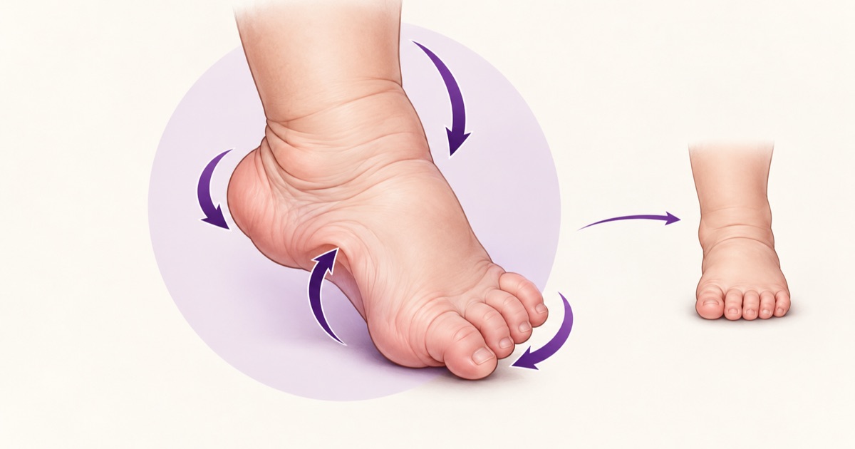
CTEV stands for congenital talipes equinovarus. The foot is held rigidly in a position where the heel is pulled up (equinus), turned inwards (varus), the forefoot is swung towards the midline (adductus) and the arch is abnormally high (cavus). It is commonly quoted as occurring in roughly 1 in 1,000 live births, about twice as often in boys, and a substantial fraction of cases, often given as a third to a half, are bilateral.
Most cases are idiopathic, meaning an isolated deformity in an otherwise normal baby. Recognised risk factors include a family history, male sex and maternal smoking. Clubfoot can also be secondary to a neuromuscular or syndromic condition, such as myelomeningocele or arthrogryposis, so a clubfoot with other joint contractures, a neurological deficit or other anomalies deserves a wider look. The deformity can often be picked up on antenatal ultrasound in the second trimester.
The Four Components: CAVE
The mnemonic CAVE names the four components, and the same order is the order of correction.
| Component | What it means | When it is corrected |
|---|---|---|
| Cavus | High arch from a plantarflexed forefoot (first ray) relative to the hindfoot | First: supinate the forefoot |
| Adductus | Forefoot deviated medially at the midfoot | Second: abduct the foot under the talus |
| Varus | Heel inverted, calcaneus turned under the talus | Second: same manoeuvre as adductus |
| Equinus | Ankle plantarflexed with a tight Achilles tendon | Last: Achilles tenotomy and final cast |
In the Ponseti method adductus and varus are corrected together, so the real sequence is cavus, then adductus and varus, then equinus.
What Is Wrong Inside the Foot
The talus is abnormal, with a short neck that is deviated medially and towards the sole. The calcaneus sits in equinus and varus, rotated inwards beneath the talus. The navicular is displaced medially, close to the medial malleolus. The soft tissues on the posteromedial side are contracted, including the tibialis posterior tendon and the ligaments around the ankle and subtalar joint.
On examination, the heel looks empty because the calcaneus is pulled up and back. There is a deep crease across the back of the heel and another along the medial arch, the foot is smaller than its partner, and the calf is thinner. The defining feature is rigidity: the foot cannot be passively corrected to a neutral position.
True Clubfoot vs Look-alikes
| Condition | Hindfoot | Forefoot | Rigidity | Course |
|---|---|---|---|---|
| True clubfoot (CTEV) | Equinus and varus | Adducted, with cavus | Rigid | Needs treatment (Ponseti) |
| Positional clubfoot | Mild equinus or varus from intrauterine position | Mildly adducted | Flexible, corrects fully | Resolves with stretching |
| Metatarsus adductus | Normal | Adducted | Usually flexible | Often resolves on its own |
| Congenital vertical talus | Rigid equinus with valgus | Dorsiflexed (rocker-bottom sole) | Rigid | Needs treatment; the opposite pattern to CTEV |
| Calcaneovalgus foot | Dorsiflexed and everted | Dorsiflexed | Flexible | Resolves on its own |
The decisive question is whether the foot corrects passively. A foot that looks turned in but corrects fully to neutral is positional and is managed with stretching. A foot that will not correct is true clubfoot.
Assessing Severity: The Pirani Score
The Pirani score grades six clinical signs, each scored 0 (normal), 0.5 (partly abnormal) or 1 (severely abnormal), for a maximum of 6. Three signs assess the hindfoot: the posterior crease, the empty heel and the rigid equinus. Three assess the midfoot: the medial crease, the curved lateral border and the coverage of the talar head. The score is recorded at the start of treatment to grade severity and repeated at each cast change to track progress. The Dimeglio score is an alternative, more detailed grading system.
The Ponseti Method: The Gold Standard
The Ponseti method is non-surgical first-line treatment and works best when started early, ideally within the first weeks of life, although it can still succeed in older infants. Treatment consists of weekly gentle manipulation followed by a long-leg plaster cast, from toes to upper thigh with the knee flexed to about 90 degrees. Correction usually takes around five to six casts.
The sequence follows CAVE:
- 1.Correct the cavus first by supinating the forefoot and lifting the first metatarsal so that the forefoot lines up with the hindfoot.
- 2.Correct adductus and varus together by abducting the foot in supination, with the head of the talus acting as the fulcrum. The foot is not pronated, and the heel is not pushed on directly.
- 3.Correct the equinus last. Most feet need a percutaneous Achilles tenotomy, done when the forefoot is corrected but the ankle still cannot dorsiflex to roughly 10 to 15 degrees. A final cast is then left on for about three weeks.
- 4.Brace to maintain correction. A foot abduction brace, the boots-and-bar type (Denis Browne bar), is worn full time for about the first three months, then at night and during naps for several years.
Why Equinus Is Corrected Last
This is the most tested logic in the topic. If the foot is forced upwards to fix equinus before the cavus and the adductus and varus are corrected, the calcaneus is still locked beneath the talus and cannot move. The force then breaks through the weakest point, the midfoot, and produces a rocker-bottom foot, a convex sole with the heel still in equinus. Fixing the forefoot and the hindfoot rotation first frees the calcaneus, and only then can the Achilles tendon be lengthened safely.
Why Casting Works, and Why Surgery Is No Longer First-Line
Ponseti treatment relies on the fact that a newborn’s ligaments, tendons and joint capsules respond unusually well to gentle, sustained stretching, which is why treatment is best started early. An earlier era used extensive surgical release of the posteromedial soft tissues as the first step. Long-term follow-up showed that a proportion of those feet became stiff and painful, and some developed early arthritis, so casting became first-line and surgery moved to a supporting role for resistant or relapsed cases. In an exam, the first step in a newborn with CTEV is serial manipulation and casting, not an operation.
Practical Details of Casting and Bracing
Each cast is changed weekly, and the foot is gently manipulated before the next cast goes on. Parents are taught to check the toes for colour, warmth and swelling, and to seek help if the cast slips, because a loose cast loses its corrective effect and can rub the skin. The abduction brace is a pair of shoes joined by a bar about as wide as the shoulders, holding the feet in outward rotation, more on the clubfoot side than on the normal side. The bar is what maintains the abduction, so wearing the shoes without the bar does not protect the correction.
Relapse and Resistant Cases
The most common cause of relapse is not wearing the brace as prescribed. Early relapse is treated with repeat casting, sometimes with a repeat tenotomy. In older children (around two and a half to three years and above) a dynamic supination deformity can develop, which is treated by transferring the tibialis anterior tendon to the lateral cuneiform.
Late, neglected or resistant feet may need a posteromedial soft tissue release. In older neglected feet, options include gradual correction with an external fixator, and triple arthrodesis is a salvage procedure once the skeleton has matured. A neglected clubfoot is recognisable because the child walks on the lateral border of the foot, with callosities over the weight-bearing area.
Associations and Practical Points
- Look for associated conditions, especially neurological problems such as spinal dysraphism and other joint contractures. An association with developmental dysplasia of the hip is described, so the hips are examined too.
- Even after successful treatment, the clubfoot side usually stays slightly smaller with a thinner calf. This is expected and is not a treatment failure.
- Explaining the reason for bracing to parents is part of treatment, because adherence is what protects the correction.
How Vignettes Are Built
Identify the components, rule out the look-alikes, then choose the first step. A two-week-old boy with a rigid foot, heel up and turned in, high arch and inward forefoot, that cannot be corrected passively, is describing CTEV, and the first step is Ponseti casting, not surgery. A foot that looks turned in but corrects fully to neutral is positional. A child who is correctly casted and tenotomised but later relapses most likely stopped wearing the brace.