Neonatal Jaundice for FMGE: Physiological vs Pathological, and the Timing Clue That Decides It
By Dr. Utsav Bhattacherjee, MBBS, MBA · 28 September 2026 · 12 min read
Neonatal jaundice questions almost always reduce to one decision: is this the normal newborn transition, or a warning sign? The fastest discriminator is not the bilirubin number but when the jaundice appeared. Learn the timing framework below, and most vignettes sort themselves before you ever look at the investigations.
Why Newborns Become Jaundiced at All
Nearly every newborn has some rise in unconjugated bilirubin in the first week, and the reasons are worth knowing as a set, because they explain both the normal pattern and what pushes it into abnormal territory.
Bilirubin production is high. Newborn red cells live for a shorter time than adult red cells, and the newborn haematocrit is high, so more haem is broken down for every kilogram of body weight.
Hepatic handling is immature. The enzyme that conjugates bilirubin, UDP-glucuronosyltransferase, has low activity in the first days of life, so the liver cannot clear the load quickly.
The gut recycles bilirubin. The newborn gut is initially sterile, feeding is only just being established, and intestinal beta-glucuronidase can deconjugate bilirubin so that it is reabsorbed. This is the enterohepatic circulation.
Put together, that is high production, slow conjugation and efficient reabsorption. Physiological jaundice is what that combination looks like when nothing else is wrong. Pathological jaundice is what happens when one of those three factors is pushed much further than normal.
Physiological Jaundice: The Pattern to Recognise
Physiological jaundice follows a predictable time course. It appears after the first 24 hours, typically on the second or third day. It peaks around day three to five in a term baby, and a little later, around day five to seven, in a preterm baby. It fades by roughly the end of the second week. The bilirubin is unconjugated, the baby is otherwise well and feeding, and the rise is gradual.
Exact cutoffs differ slightly between textbooks and guidelines, so it is safer to learn the pattern than a single number: late onset, slow rise, peak in the first week, spontaneous resolution, and a well baby. Any departure from that pattern, especially early onset or a fast rise, moves the case towards pathological.
Pathological Jaundice: The Red Flags
Jaundice is considered pathological when it breaks the physiological pattern. The commonly cited red flags are:
- Onset within the first 24 hours of life. This alone makes it pathological until proven otherwise.
- A rapid rise in serum bilirubin, commonly cited as more than about 5 mg/dL per day.
- Raised conjugated (direct) bilirubin. This is never a normal newborn finding.
- Persistence beyond about two weeks in a term baby, or about three weeks in a preterm baby.
- Signs of illness, such as poor feeding, lethargy, temperature instability, pale stools or dark urine.
If you remember only one of these for the exam, remember the first. Jaundice in the first 24 hours is never physiological, and that timing points straight to haemolysis or infection.
The Timing Framework: Causes by Day of Onset
The most useful way to organise the causes is by the day the jaundice appears.
| Onset | Think of | Why |
|---|---|---|
| Within 24 hours | Rh or ABO incompatibility, G6PD deficiency, congenital infection | Red cells are being destroyed from birth |
| Day 2 to 3 | Physiological jaundice; also sepsis, polycythaemia, cephalhaematoma | Normal transition, or an extra bilirubin load from another source |
| Day 3 to 7 | Sepsis, breastfeeding jaundice, G6PD deficiency, ongoing haemolysis | Poor intake and rising reabsorption, or infection |
| After the first week | Breast milk jaundice, hypothyroidism, urinary tract infection, galactosaemia | Causes of prolonged unconjugated jaundice |
| Beyond 2 weeks with dark urine and pale stools | Biliary atresia, neonatal hepatitis, choledochal cyst | Conjugated hyperbilirubinaemia (cholestasis) |
Rh Versus ABO Incompatibility
Two isoimmune causes dominate early-onset jaundice, and the differences between them are a favourite comparison question.
Rh incompatibility occurs when an Rh-negative mother carries an Rh-positive baby and has been sensitised, usually by a previous Rh-positive pregnancy, delivery, miscarriage or transfusion. The first affected baby is usually spared, because sensitisation typically happens at delivery, so the disease tends to worsen with each subsequent pregnancy. The direct Coombs test is strongly positive, anaemia can be severe, and in the worst cases the baby develops hydrops fetalis.
ABO incompatibility usually occurs when the mother is group O and the baby is group A or B. It can affect the first pregnancy, because group O mothers already carry naturally occurring IgG anti-A and anti-B antibodies that cross the placenta. It is generally milder, the direct Coombs test is often only weakly positive or even negative, and spherocytes on the peripheral smear are a characteristic clue.
| Feature | Rh incompatibility | ABO incompatibility |
|---|---|---|
| Mother and baby | Rh-negative mother, Rh-positive baby | Group O mother, group A or B baby |
| First pregnancy affected? | Usually not | Yes, it can be |
| Severity | Often severe; hydrops possible | Usually mild to moderate |
| Direct Coombs test | Strongly positive | Weak or negative |
| Spherocytes on smear | Not typical | Common |
| Prevention | Anti-D immunoglobulin for Rh-negative mothers | No specific prophylaxis |
Rh disease is preventable. Anti-D immunoglobulin given to an Rh-negative mother after delivery of an Rh-positive baby (within 72 hours) stops her from becoming sensitised in the first place, which is why the disease is now uncommon where this is routinely done.
Breastfeeding Jaundice vs Breast Milk Jaundice
These two names sound alike and mean very different things, so examiners like them.
Breastfeeding jaundice appears in the first week and is caused by inadequate intake. The baby is not getting enough milk, so there is dehydration, less stool and more reabsorption of bilirubin from the gut. The fix is better feeding: lactation support, more frequent feeds, and checking weight loss.
Breast milk jaundice appears later, usually after the first week, and can last several weeks. The baby is thriving and feeding well, the bilirubin is unconjugated, and the cause is thought to be substances in the milk that increase bilirubin reabsorption. It is benign and is a diagnosis of exclusion. The classic exam trap is that breastfeeding should generally be continued, not stopped.
Kernicterus: Why Unconjugated Bilirubin Is Dangerous
Unconjugated bilirubin is fat-soluble and can cross the blood-brain barrier. When levels are high enough, or the baby’s defences are weakened, it deposits in the basal ganglia (especially the globus pallidus), brainstem nuclei and cerebellum. This is bilirubin encephalopathy, and its chronic form is called kernicterus.
The acute phase begins with lethargy, poor sucking and hypotonia, and can progress to hypertonia, arching of the back (opisthotonos), a high-pitched cry and seizures. The chronic phase leaves permanent damage: athetoid cerebral palsy, sensorineural hearing loss, upward gaze palsy and dental enamel dysplasia.
Risk rises with prematurity, haemolysis, sepsis, acidosis and low albumin. Certain drugs, such as sulfonamides and ceftriaxone, compete with bilirubin for albumin binding and are avoided in the jaundiced newborn for this reason.
Assessment and Investigations
Yellow discolouration spreads from head to toe as bilirubin rises, and palm and sole involvement suggests a high level. This head-to-toe progression (Kramer’s rule) is only an approximation and becomes unreliable in darker skin and once phototherapy has begun, so confirm with a serum bilirubin.
A sensible first set of tests is total and direct bilirubin, blood group and Rh type of mother and baby, a direct Coombs test, a full blood count with reticulocyte count and smear, and G6PD testing. Add a sepsis screen if the baby looks unwell, and thyroid function tests plus a urine test for reducing substances (galactosaemia) if the jaundice is prolonged.
Treatment: Phototherapy and Exchange Transfusion
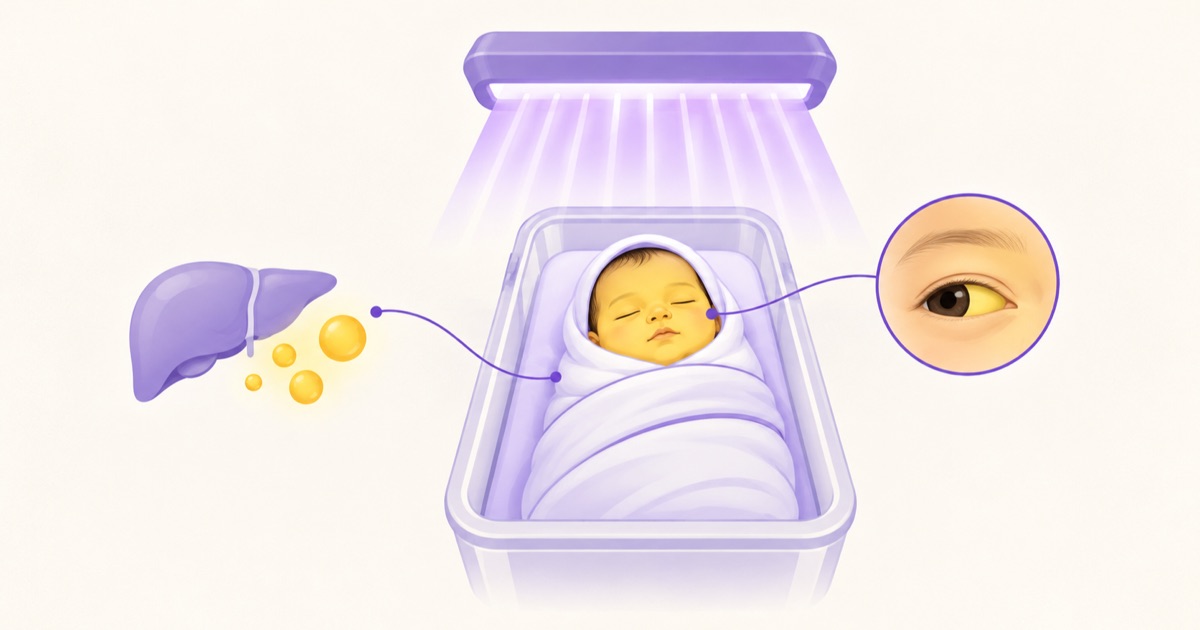
Phototherapy uses blue-green light, roughly in the 460 to 490 nanometre range. It converts bilirubin in the skin into water-soluble photoisomers that are excreted in bile and urine without needing conjugation, which is why it works despite the immature liver. The baby is exposed with eyes covered, kept hydrated and monitored for temperature. Side effects include loose stools, dehydration, a transient rash and temperature instability. In babies with conjugated hyperbilirubinaemia, phototherapy can cause bronze baby syndrome, a grey-brown discolouration of the skin.
Exchange transfusion is reserved for severe hyperbilirubinaemia that is not responding to intensive phototherapy, or for any baby showing signs of acute bilirubin encephalopathy. It removes bilirubin, antibody-coated red cells and circulating antibodies. Its risks include electrolyte disturbances such as hypocalcaemia, low platelets, infection and catheter-related complications. In isoimmune haemolytic disease, intravenous immunoglobulin can reduce the need for exchange transfusion.
Treatment thresholds come from charts based on age in hours, gestation and risk factors, so learn the logic rather than a single number.
Conjugated Jaundice Is Never Physiological
Raised conjugated (direct) bilirubin always needs investigation. The classic warning pattern is jaundice persisting beyond two weeks with dark urine and pale, clay-coloured stools, which points to cholestasis. Causes include biliary atresia, neonatal hepatitis, choledochal cyst, galactosaemia and cholestasis linked to parenteral nutrition.
Biliary atresia is the one that cannot be missed, because outcomes depend on timing. The Kasai portoenterostomy gives the best results when it is done early, ideally within the first two months of life. A baby with prolonged jaundice and pale stools should be investigated urgently, not reassured.
Prolonged unconjugated jaundice has its own important cause. Hypothyroidism should be considered, particularly when there is lethargy, constipation, a large fontanelle or an enlarged tongue.
How Vignettes Are Built
Work through the same steps every time: age at onset, conjugated or unconjugated, evidence of haemolysis (blood groups, Coombs test, smear), evidence of infection, and feeding pattern.
A baby yellow at 18 hours with a group O mother, a group A baby and a weakly positive Coombs test is describing ABO incompatibility. A thriving, exclusively breastfed three-week-old with unconjugated jaundice and normal stool colour is describing breast milk jaundice. The same baby with pale stools and dark urine is a different problem entirely: cholestasis until proven otherwise.