Trachoma for FMGE: WHO Grading, Complications, and the SAFE Strategy
By Dr. Utsav Bhattacherjee, MBBS, MBA · 28 September 2026 · 12 min read
Trachoma questions test three connected ideas: how a repeated childhood eye infection ends in blindness, how the disease is classified (the MacCallan stages and the WHO simplified grading), and how it is controlled (the SAFE strategy). Keep the chain from infection to blindness in mind, and the classification and the control measures both make sense.
The Organism and How It Spreads
Trachoma is caused by Chlamydia trachomatis, serotypes A, B, Ba and C. The genital and inclusion conjunctivitis serotypes are D to K, and L1 to L3 cause lymphogranuloma venereum, so knowing that A to C means trachoma is a reliable exam point. Chlamydiae are obligate intracellular bacteria with two forms: the elementary body, which is infectious, and the reticulate body, which multiplies inside the cell and forms the intracytoplasmic inclusions seen on staining.
Spread is from eye to eye, through fingers, shared cloths and towels, and eye-seeking flies. Overcrowding, poor sanitation, scarce water and poor facial hygiene drive transmission, and young children are the main reservoir of active infection.
From Infection to Blindness: The Chain
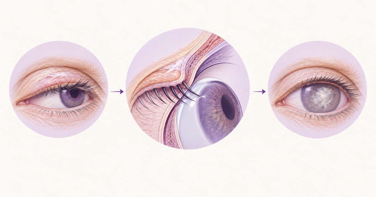
The blindness in trachoma does not come from a single infection. It comes from repeated infections over years, and the sequence is worth learning as a chain:
- 1.Repeated chlamydial infection causes chronic follicular conjunctivitis.
- 2.Chronic inflammation leads to scarring of the upper tarsal conjunctiva.
- 3.The scar contracts and turns the lid margin inwards, causing entropion.
- 4.The lashes now rub against the eye, which is trichiasis.
- 5.The lashes abrade the cornea, causing ulceration and corneal opacity.
- 6.Corneal opacity leads to irreversible blindness.
This is why the active infection is treated with antibiotics but the blinding stage, trichiasis, is treated with surgery. Women are affected more often than men, largely through caregiving contact with infected children.
Why Repeated Infection Matters
A single episode of chlamydial conjunctivitis rarely blinds. Scarring is driven by the chronic inflammation and immune response that repeated infections keep switching on, which is why children who are reinfected many times are the ones who develop scarring in adult life. It also explains why treating one child without changing the household or community environment often fails: reinfection restarts the cycle.
Clinical Features
In active disease, which is seen mainly in children, the upper tarsal conjunctiva shows follicles and papillary hypertrophy, and the cornea may show superficial keratitis and pannus. Healing leaves characteristic scars.
| Sign | What it means |
|---|---|
| Follicles on the upper tarsal conjunctiva | Hallmark of active disease |
| Papillary hypertrophy | Intense inflammation that can hide the deep tarsal vessels |
| Pannus | Superficial vascularisation of the cornea, seen first at the upper margin |
| Herbert’s pits | Healed limbal follicles, seen as small depressions; a hallmark of past trachoma |
| Arlt’s line | A linear scar across the upper tarsal conjunctiva |
| Trichiasis | Lashes turned inwards, touching the cornea |
The MacCallan Classification
MacCallan divides trachoma into four stages:
- Stage I (incipient): immature follicles and early keratitis.
- Stage II (established): mature follicles and papillary hypertrophy, often described as IIa (follicular) and IIb (papillary).
- Stage III (cicatrising): scarring appears as the follicles heal.
- Stage IV (healed): the disease is no longer active, but the scars and their consequences remain.
The WHO Simplified Grading
The WHO simplified system is the one used for community surveys and is the most frequently tested. It has five signs, and the first two indicate active disease while the last three indicate scarring and its consequences.
| Grade | Meaning | Active disease or sequelae |
|---|---|---|
| TF | Trachomatous inflammation, follicular: five or more follicles on the upper tarsal conjunctiva | Active |
| TI | Trachomatous inflammation, intense: thickening that hides more than half of the normal deep tarsal vessels | Active |
| TS | Trachomatous scarring: easily visible scars on the tarsal conjunctiva | Sequela |
| TT | Trachomatous trichiasis: at least one lash rubbing on the eyeball | Sequela |
| CO | Corneal opacity: easily visible opacity over at least part of the pupil margin | Sequela |
For control programmes, TF prevalence in children aged one to nine is used to decide where mass treatment is needed, and TT in adults is used to plan surgery. The commonly cited elimination targets are TF below 5% in children aged one to nine, and TT unknown to the health system below 0.2% in adults aged fifteen and above.
Diagnosis
Trachoma is diagnosed clinically. The WHO definition requires at least two of four signs: lymphoid follicles on the upper tarsal conjunctiva, typical conjunctival scarring, limbal follicles or their sequela (Herbert’s pits), and vascular pannus. Laboratory confirmation is not needed for routine practice but includes Giemsa-stained conjunctival scrapings showing intracytoplasmic inclusions (Halberstaedter-Prowazek bodies), immunofluorescence, and nucleic acid amplification tests.
Treatment
Active trachoma is treated with a single dose of oral azithromycin, given at about 20 mg/kg up to a 1 g maximum, which is also the drug used for mass treatment of communities. Topical tetracycline 1% eye ointment, applied twice daily for about six weeks, is the traditional alternative.
Antibiotics do not fix trichiasis. The definitive treatment is surgery, and the WHO-recommended operation is bilamellar tarsal rotation, which rotates the lid margin outwards so the lashes no longer touch the eye. Epilation is only a temporary measure. Established corneal opacities may need corneal transplantation.
Mass Treatment in Practice
Because infection circulates within families and communities, control programmes treat whole communities rather than individual cases. In districts where TF prevalence in children is above the WHO thresholds, everyone in the community is offered azithromycin, usually once a year for several years. Trichiasis patients are found and offered surgery through outreach camps and district hospitals, and surveys of children aged one to nine are repeated to see whether TF has fallen below the elimination target.
The SAFE Strategy
SAFE is the WHO-recommended control strategy, and each letter targets a different part of the disease.
| Letter | Component | What it does |
|---|---|---|
| S | Surgery | Treats trichiasis to prevent blindness in people who already have late disease |
| A | Antibiotics | Reduces the reservoir of active chlamydial infection |
| F | Facial cleanliness | Cuts eye-to-eye and fly-borne transmission |
| E | Environmental improvement | Water, sanitation and fly control, which prevent reinfection over the long term |
Note the logic: S protects people who are already at risk, A treats infection now, and F and E stop it coming back. Antibiotics alone do not eliminate trachoma, because without F and E reinfection follows.
Complications and Sequelae
The main complications follow the chain above and can be grouped by site. Lids: trichiasis, entropion and ptosis. Conjunctiva: scarring, xerosis (dry eye from destruction of conjunctival goblet cells) and symblepharon. Cornea: ulceration, opacity and ectasia. Lacrimal system: scarring of the ducts and chronic dacryocystitis.
Trachoma vs Inclusion Conjunctivitis
Both are caused by Chlamydia trachomatis, which is why they get compared.
| Feature | Trachoma | Inclusion conjunctivitis |
|---|---|---|
| Serotypes | A, B, Ba, C | D to K |
| Transmission | Eye to eye, flies, poor hygiene | Genital to eye in adults; birth canal in neonates |
| Setting | Crowded, low-resource communities | Sexually active adults and newborns |
| Course | Chronic, repeated, ends in scarring | Usually self-limited, little or no scarring |
| Treatment | Azithromycin or tetracycline; SAFE strategy | Systemic azithromycin or doxycycline; treat partners |
India’s Trachoma Story
Trachoma was once a leading cause of blindness in India. The National Trachoma Control Programme began in 1963 and was later folded into the national programme for control of blindness. On 8 October 2024 the WHO validated India as having eliminated trachoma as a public health problem. Trachoma-related blindness is reported to have fallen from about 5% of all blindness in 1971 to under 1%.
Elimination as a public health problem does not mean the disease has vanished. It means the burden is below WHO thresholds, and the classification, grading and management of trachoma remain standard examination material.
How Vignettes Are Built
Look for where the patient is on the chain. A child with follicles on the upper tarsal conjunctiva and no scarring has active disease (TF), so the answer is azithromycin. An adult woman from a low-resource community with lashes rubbing the cornea (TT) needs bilamellar tarsal rotation, not more antibiotics. An adult with a visible corneal scar over the pupil margin is CO, the blinding stage, and the question is usually about the grade. Serotype clues settle trachoma versus inclusion conjunctivitis questions immediately: A to C is trachoma, D to K is inclusion conjunctivitis.