Types of Hernias for FMGE: The One Landmark That Answers Every Groin Hernia Question
By Dr. Utsav Bhattacherjee, MBBS, MBA · 4 September 2026 · 11 min read
Almost every groin hernia question comes down to one anatomical relationship: where the hernia sac sits relative to the inferior epigastric vessels. Get that landmark fixed in mind, and indirect, direct, and femoral hernias stop being three things to memorize separately and become three positions around a single fixed point.
The Landmark: Inferior Epigastric Vessels
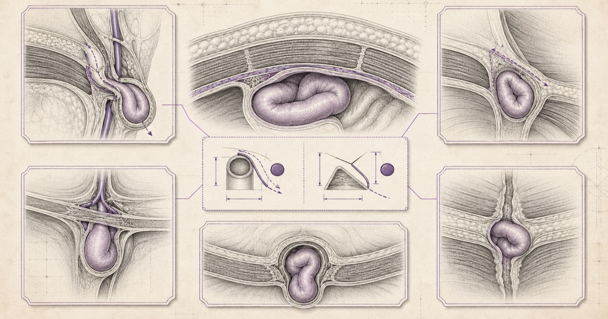
The inferior epigastric vessels run vertically along the posterior abdominal wall and serve as the dividing line examiners build groin hernia questions around. A hernia sac emerging lateral to these vessels is indirect. A hernia sac emerging medial to these vessels, through the floor of Hesselbach’s triangle, is direct. This single relationship is worth over-learning before anything else in this topic, because it’s the fact that actually resolves most vignettes.
Indirect Inguinal Hernia
Indirect inguinal hernia is congenital in origin — it follows the path of the processus vaginalis, the embryological tract along which the testis descends into the scrotum during development. When this tract fails to obliterate properly after descent, it leaves a potential pathway for abdominal contents to herniate through. The hernia sac exits through the deep inguinal ring, travels through the inguinal canal alongside the spermatic cord, and can potentially extend all the way into the scrotum in its more advanced form.
Because it’s congenital, indirect inguinal hernia is the type most commonly seen in infants and young patients, though it can present at any age if the anatomical predisposition was always present but only became symptomatic later. It is also, overall, the most common type of hernia across all ages and both sexes.
Direct Inguinal Hernia
Direct inguinal hernia is acquired, arising from a genuine weakness in the transversalis fascia within Hesselbach’s triangle — the anatomical space bounded by the inferior epigastric vessels laterally, the rectus abdominis muscle medially, and the inguinal ligament inferiorly. Unlike the indirect type, there’s no embryological remnant driving this — it develops from chronic increases in intra-abdominal pressure acting on a wall that has weakened over time.
This makes direct inguinal hernia predominantly a condition of older men, and a useful clinical clue: because the defect is a broad area of fascial weakness rather than a discrete tract, direct hernias tend to reduce easily and rarely strangulate compared to the other two types on this list — a detail worth remembering when a vignette is testing which hernia type carries genuine surgical urgency.
Femoral Hernia
Femoral hernia takes a different path entirely — below the inguinal ligament, through the femoral canal, medial to the femoral vein. It’s more common in women, largely attributed to differences in pelvic anatomy that create a comparatively wider femoral canal.
The single most important exam fact about femoral hernias: they carry the highest risk of strangulation of the three groin hernia types, because the femoral canal is a rigid, narrow space with little room for the herniated tissue to move without its blood supply being compressed. A femoral hernia presenting with sudden pain and a tender, irreducible groin mass is a surgical emergency in a way a reducible direct inguinal hernia typically isn’t.
Indirect vs Direct vs Femoral: Side by Side
| Feature | Indirect Inguinal | Direct Inguinal | Femoral |
|---|---|---|---|
| Origin | Congenital (patent processus vaginalis) | Acquired (fascial weakness) | Acquired |
| Position relative to inferior epigastric vessels | Lateral | Medial | Below inguinal ligament |
| Exit point | Deep inguinal ring | Hesselbach’s triangle | Femoral canal |
| Typical demographic | Younger patients, infants | Older men | Women |
| Strangulation risk | Moderate | Low | High |
Named Hernias Worth Knowing Separately
- Richter’s hernia — only part of the bowel circumference (not the full lumen) becomes trapped in the defect. This is a genuinely dangerous pattern to recognise: because the bowel lumen isn’t fully obstructed, the patient may not show classic signs of bowel obstruction even while the trapped segment is strangulating and at real risk of perforation.
- Littre’s hernia — a hernia sac containing a Meckel’s diverticulum. The name is specific to this exact combination and is a common "match the named hernia to its definition" exam pairing.
- Spigelian hernia — protrudes through the linea semilunaris, along the lateral edge of the rectus abdominis muscle, at a point below the umbilicus. It’s notable for being easy to miss on exam, since it often stays within the abdominal wall layers without an obvious external bulge.
- Obturator hernia — passes through the obturator canal, and is classically associated with elderly, thin women. The Howship-Romberg sign — pain radiating down the inner thigh, from compression of the obturator nerve — is the specific finding examiners attach to this hernia, and its presence in a vignette is usually a deliberate signal rather than incidental detail.
The Inguinal Canal: Boundaries Worth Memorising Separately
The canal itself is tested as a standalone anatomy question often enough to be worth learning apart from the hernias that pass through it. Its floor is the inguinal ligament, its roof the arching fibres of internal oblique and transversus abdominis, its anterior wall the external oblique aponeurosis (reinforced laterally by internal oblique), and its posterior wall the transversalis fascia, reinforced medially by the conjoint tendon. The pattern worth holding onto is that the anterior wall is reinforced laterally while the posterior wall is reinforced medially — which is precisely why an indirect hernia pushes through laterally and a direct one through the weaker medial posterior wall.
Contents differ by sex, and that difference is itself a question. In males the canal transmits the spermatic cord; in females, the round ligament of the uterus. Both carry the ilioinguinal nerve, and its course is clinically load-bearing: it is the nerve most at risk during open inguinal hernia repair, and injury to it produces numbness or persistent neuropathic pain over the medial thigh and scrotum or labium. Chronic post-herniorrhaphy groin pain is common enough that a vignette describing persistent burning pain in that distribution after a repair is usually pointing at ilioinguinal nerve entrapment or injury rather than recurrence.
Recurrence is the other post-repair complication worth separating from that picture. A recurrent hernia presents as a reappearing bulge at the operative site, reducible at first, rather than as pain in a nerve distribution — so the two are distinguished by what the patient reports, not by imaging.
Beyond the Groin: Umbilical and Incisional Hernias
Not every hernia FMGE tests is a groin hernia, and two abdominal wall hernias are worth knowing on their own terms.
Umbilical hernia has a genuinely useful congenital-versus-acquired split, similar in spirit to the indirect/direct distinction above. The congenital form is common in infants, caused by incomplete closure of the umbilical ring after birth, and the reassuring exam fact is that the large majority resolve spontaneously by 2–4 years of age without intervention — surgical repair is generally reserved for defects that persist beyond that window or are unusually large from the start. The acquired form in adults is a different problem entirely, driven by chronic increases in intra-abdominal pressure — obesity, pregnancy, and ascites are the three classic associations — and unlike the pediatric form, adult umbilical hernias don’t resolve on their own and are managed surgically once identified.
Incisional hernia develops through a previous surgical scar, at a site where the fascial layer closed during the original operation has since failed. Risk factors cluster around anything that impairs normal wound healing or adds mechanical stress to the closure: wound infection at the original surgical site, obesity, poor nutritional status, chronic cough, and technical factors in how the original closure was performed. A hernia appearing at the site of a prior abdominal surgery, sometimes months or years later, is describing an incisional hernia by definition — the surgical history in the vignette is doing most of the diagnostic work.
What Actually Determines Surgical Urgency
The clinical distinction that matters most isn’t which named type a hernia is, but whether it’s reducible, incarcerated, or strangulated. A reducible hernia can be pushed back into the abdominal cavity and carries no immediate urgency. An incarcerated hernia is stuck and cannot be reduced, but blood supply to the trapped tissue is still intact — this needs prompt attention but isn’t yet a true emergency. A strangulated hernia has lost its blood supply, and the trapped tissue is at risk of necrosis within hours — this is a surgical emergency regardless of which named type of hernia is involved. Femoral hernias reach strangulation more often simply because their anatomy makes incarceration more likely in the first place, not because strangulation itself works any differently once it occurs.