Types of Shock for FMGE: The Hemodynamic Table That Answers the Question Every Time
By Dr. Utsav Bhattacherjee, MBBS, MBA · 4 September 2026 · 10 min read
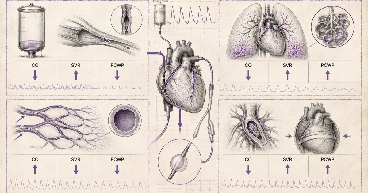
Shock questions on FMGE are almost always solved the same way: identify the cardiac output, systemic vascular resistance, and preload pattern described in the vignette, and match it against the table below. The clinical scenario tells you the cause; the hemodynamic numbers confirm the type.
What Shock Actually Means
Shock is a state of inadequate tissue perfusion relative to metabolic demand — not necessarily low blood pressure, though the two often coexist. The underlying problem can come from three different places: not enough volume in the system (hypovolemic), a pump that isn’t working (cardiogenic), vessels that have lost their tone and are pooling blood in the wrong places (distributive), or a mechanical blockage preventing blood from flowing where it needs to go (obstructive). Every named type of shock below is a variation on one of these four root causes.
Hypovolemic Shock
Hypovolemic shock results from a genuine loss of intravascular volume — hemorrhage is the classic cause, but severe burns, prolonged vomiting or diarrhea, and third-spacing in conditions like pancreatitis all produce the same underlying picture. Cardiac output falls because there simply isn’t enough volume returning to the heart to fill it properly, and the body compensates with increased systemic vascular resistance — peripheral vasoconstriction, in an attempt to maintain blood pressure despite the falling output. Central venous pressure (preload) falls as well, since the volume that would normally fill the right heart is what’s missing in the first place.
Clinically, this produces the classic "cold shock" picture: cool, clammy extremities from peripheral vasoconstriction, tachycardia as a reflex compensatory response, and a narrowing pulse pressure as the body works to maintain perfusion with less volume to work with.
Cardiogenic Shock
Cardiogenic shock is a pump failure — the heart itself cannot generate adequate output despite adequate volume being available to it. Myocardial infarction is the classic cause, but severe arrhythmias, acute valve rupture, and myocarditis can all produce the same picture. Cardiac output falls, just as in hypovolemic shock, and systemic vascular resistance rises for the same compensatory reason — but the critical distinguishing feature is that central venous pressure (preload) rises rather than falls, because the failing heart can’t move forward the volume that’s arriving at it, and blood backs up behind the failing pump.
This CVP distinction is the single most tested differentiator between hypovolemic and cardiogenic shock — two conditions that can otherwise look similar at the bedside (both present as "cold shock," both show low output and high resistance), but diverge completely on this one hemodynamic parameter.
Distributive Shock
Distributive shock is a fundamentally different mechanism from the two above — the problem isn’t volume or pump function, it’s that the blood vessels themselves have lost normal tone and are inappropriately dilated, pooling blood away from where it’s needed. This produces a hemodynamic pattern that’s the near-opposite of hypovolemic and cardiogenic shock in its early phase: systemic vascular resistance falls rather than rises, and cardiac output is often normal or even elevated, at least initially, as the heart tries to compensate for the fallen resistance by pumping harder and faster.
Septic shock is the most common form of distributive shock, caused by the systemic inflammatory response to infection triggering widespread vasodilation. It has a genuinely useful early-versus-late distinction: the early ("warm") phase shows warm, flushed extremities from vasodilation, with a bounding pulse and low SVR — the opposite of the cold, clammy picture in hypovolemic or cardiogenic shock. As septic shock progresses and compensatory mechanisms fail, it can transition to a "cold" phase that becomes harder to distinguish from other shock types on exam alone.
Anaphylactic shock is IgE-mediated, triggered by re-exposure to an allergen the immune system has already been sensitized to. Mast cell degranulation releases histamine and other mediators, causing rapid, severe vasodilation and increased vascular permeability — the onset is typically much faster than septic shock, often within minutes of exposure, and is frequently accompanied by urticaria, angioedema, and bronchospasm that point clearly toward the diagnosis.
Neurogenic shock follows a high spinal cord injury (classically above T6) that disrupts the sympathetic nervous system’s normal vasoconstrictor tone below the level of injury. The vasodilation itself matches the distributive pattern, but the detail that separates neurogenic shock from every other type on this list is the heart rate: bradycardia, not the reflex tachycardia seen in hypovolemic, cardiogenic, and most septic shock. Unopposed vagal (parasympathetic) tone, with the sympathetic chain disrupted, is what produces this — a vignette describing hypotension with bradycardia after a spinal injury is describing neurogenic shock specifically, not a generic distributive picture.
Obstructive Shock
Obstructive shock is a mechanical problem — something is physically blocking blood flow, even though both the pump and the vasculature are otherwise intact. Tension pneumothorax, cardiac tamponade, and massive pulmonary embolism are the three classic causes, and each obstructs flow at a different point, but all three produce a shared pattern: cardiac output falls, and preload is typically elevated on the venous side proximal to the obstruction — blood backing up behind the blockage, similar in principle to the backup seen in cardiogenic shock, but from a mechanical cause rather than pump failure itself.
Cardiac tamponade specifically presents with Beck’s triad — hypotension, distended neck veins (elevated JVP), and muffled heart sounds — a classically tested triad that points directly to fluid compressing the heart from outside. Tension pneumothorax presents with absent breath sounds on the affected side, tracheal deviation away from the affected side, and distended neck veins from impaired venous return — a genuine emergency requiring immediate needle decompression rather than imaging confirmation first.
The Hemodynamic Table
| Type | Cardiac Output | SVR | Preload (CVP) |
|---|---|---|---|
| Hypovolemic | Decreased | Increased | Decreased |
| Cardiogenic | Decreased | Increased | Increased |
| Distributive (septic, early) | Normal/Increased | Decreased | Variable |
| Distributive (anaphylactic) | Variable, often decreased | Decreased | Decreased |
| Distributive (neurogenic) | Decreased (with bradycardia) | Decreased | Decreased |
| Obstructive | Decreased | Increased | Increased (proximal to obstruction) |
The single most efficient way to work through a shock vignette: read the heart rate first (bradycardia narrows almost immediately to neurogenic shock, since everything else on this list produces reflex tachycardia), then match the CVP direction against hypovolemic (down) versus cardiogenic or obstructive (up), then use the clinical scenario to separate cardiogenic from obstructive when both show a similar hemodynamic picture.
Why Lactate Matters Across Every Type
Regardless of which of the four mechanisms is driving it, shock ultimately means cells aren’t getting enough oxygen to meet demand, and cells forced into anaerobic metabolism produce lactate as a byproduct. This makes serum lactate a genuinely useful marker that cuts across the whole classification above — an elevated lactate confirms that perfusion is inadequate at the tissue level, independent of which hemodynamic pattern is present, and it’s often abnormal before blood pressure itself drops far enough to be obviously concerning.
Lactate is equally useful on the way back out of shock: a falling lactate over serial measurements is one of the better available indicators that resuscitation is actually working at the tissue level, not just that blood pressure numbers have improved. A vignette describing a patient whose blood pressure has normalized but whose lactate remains elevated is describing incomplete resuscitation, not a resolved case — the numbers on the monitor and the numbers on the metabolic panel don’t always move together, and treating the blood pressure alone without confirming lactate clearance is a recognized pitfall in shock management.
Quick Clinical Differentiators
- Cold, clammy skin points toward hypovolemic or cardiogenic shock — both are low-output states with compensatory vasoconstriction.
- Warm, flushed skin points toward early distributive shock — the vasodilation itself is often visible at the bedside before the numbers confirm it.
- Bradycardia instead of tachycardia is the single fastest route to neurogenic shock, since it breaks the pattern every other type on this list follows.
- Beck’s triad (hypotension, distended neck veins, muffled heart sounds) means cardiac tamponade specifically, not obstructive shock generically.
- Absent breath sounds with tracheal deviation means tension pneumothorax, and management (needle decompression) shouldn’t wait for imaging.