Ziehl-Neelsen Staining for FMGE: Why Mycobacteria Need a Different Stain Entirely
By Dr. Utsav Bhattacherjee, MBBS, MBA · 23 September 2026 · 12 min read
Ziehl-Neelsen staining questions are built around one core fact: it exists specifically because Mycobacteria have a cell wall Gram staining cannot penetrate reliably, and the entire technique is designed around forcing a stain through that waxy barrier and then proving it stays put. Understand that one design goal, and every step of the procedure explains itself.
Why This Stain Exists at All
Mycobacteria, including the tuberculosis-causing species, have a cell wall rich in mycolic acids — long-chain fatty acids that make the wall waxy and highly resistant to standard aqueous dyes like those used in Gram staining. This is exactly why Mycobacteria are neither reliably Gram-positive nor Gram-negative in practice; the stain simply doesn’t penetrate the wall consistently enough to classify them that way. Ziehl-Neelsen staining was developed specifically to force a dye through this resistant wall and then prove, through the technique’s core step, that the organism genuinely retains it.
The Three-Step Procedure, and Why Each Step Exists
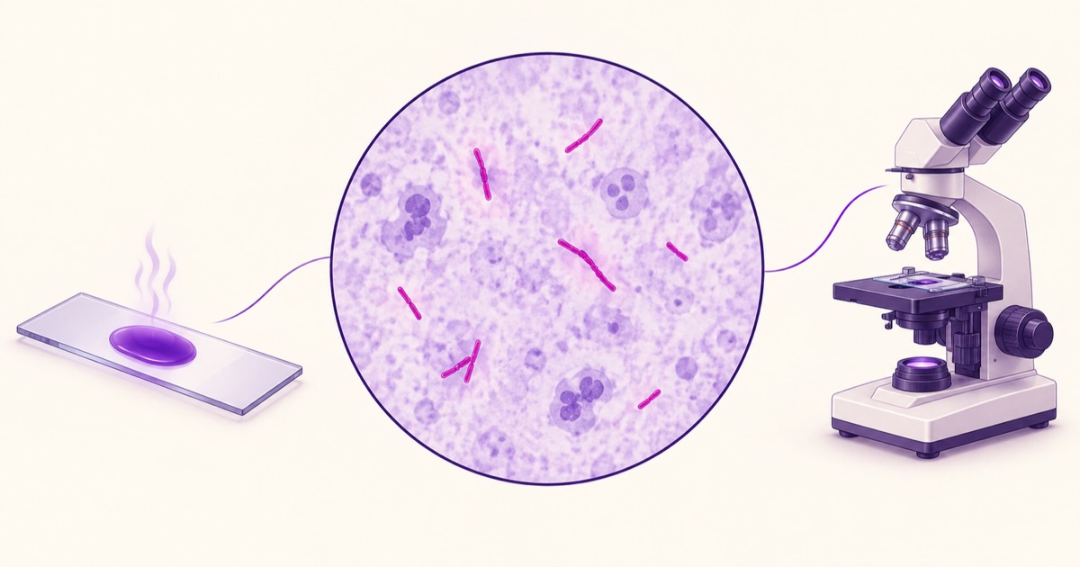
Carbol fuchsin, the primary stain, is applied with heat — traditionally by passing a flame beneath the slide until the stain steams, though some modern protocols use a chemical alternative to heat instead. The heat is not incidental; it’s what drives the stain through the waxy mycolic acid layer that would otherwise block it entirely at room temperature.
Acid-alcohol is then applied as the decolorizing agent. This is the step that gives the technique its name and its diagnostic power: an ordinary organism, lacking a mycolic acid wall, loses the carbol fuchsin stain immediately under this acid-alcohol wash. A genuinely acid-fast organism resists this decolorization specifically because the mycolic acid wall that made staining difficult in the first place now works in reverse, trapping the dye against the same acid-alcohol wash that would strip it from anything else.
Methylene blue, the counterstain, is applied last. Organisms that lost the carbol fuchsin during decolorization pick up this blue counterstain instead, while acid-fast organisms remain their original red-pink color, now standing out clearly against a blue background.
How AFB Smears Are Actually Graded
A positive Ziehl-Neelsen smear isn’t just reported as positive or negative — it’s graded based on the number of acid-fast bacilli (AFB) seen per field under the microscope, and this grading carries real clinical weight. A smear showing 1-9 AFB per 100 fields is typically reported as scanty. Progressively higher counts move through 1+ (1-9 AFB per field), 2+ (1-9 AFB per field across ten fields, roughly), and 3+ (more than 9 AFB per field) — the exact cutoffs vary slightly by protocol, but the underlying principle holds across all of them: a higher bacillary load on smear correlates with higher infectivity, which is part of why smear grading factors into both treatment monitoring and infection control decisions, not just initial diagnosis.
Other Acid-Fast Organisms Worth Knowing
Mycobacterium tuberculosis is the classic organism this stain is built around, but it’s worth knowing that acid-fastness isn’t unique to Mycobacteria alone. Nocardia species show partial acid-fastness, typically requiring a weaker decolorizing agent than standard Ziehl-Neelsen to demonstrate this property — a detail worth knowing since Nocardia can otherwise resemble other branching organisms on a smear. Cryptosporidium and Cyclospora, both intestinal parasites rather than bacteria, are also acid-fast and are specifically diagnosed using a modified Ziehl-Neelsen stain on stool samples, extending this same core technique well beyond mycobacterial disease into parasitology.
The Modified (Cold) Kinyoun Method
The Kinyoun method achieves the same acid-fast staining principle without the heating step, using a higher concentration of carbol fuchsin and phenol to force stain penetration at room temperature instead. This is a genuinely practical variation worth knowing: it removes the need for a heat source (relevant in settings without reliable access to a flame or heating element) at the cost of requiring a more concentrated stain solution, but the underlying acid-fast principle being tested is identical to conventional Ziehl-Neelsen.
Why Sample Quality Matters as Much as the Stain Itself
For pulmonary tuberculosis specifically, sputum sample quality genuinely affects diagnostic yield independent of how well the staining technique itself is performed. An early-morning sample, collected after an overnight accumulation of secretions, generally contains a higher bacillary concentration than a sample collected later in the day. This is why standard TB diagnostic protocols typically request multiple sputum samples across different collection times rather than relying on a single specimen — a negative smear from a poor-quality sample doesn’t reliably rule out disease the way a well-collected, well-stained positive smear reliably confirms it.
Why a Negative Smear Doesn’t Rule Out Tuberculosis
It’s worth holding onto a genuinely important limitation: a negative Ziehl-Neelsen smear does not exclude tuberculosis, since the bacillary load needs to reach a minimum threshold, roughly 5,000 to 10,000 organisms per millilitre of sputum, before the organism becomes reliably visible on a stained smear at all. Paucibacillary disease, or disease in a site other than the lungs, can genuinely produce a negative smear despite active infection, which is exactly why culture and molecular testing remain necessary alongside smear microscopy rather than being replaced by it.
Fluorescence Microscopy as a Modern Alternative
Auramine-rhodamine fluorescence staining works on the same acid-fast principle but uses a fluorescent dye instead of carbol fuchsin, examined under a fluorescence microscope rather than standard light microscopy. The genuine advantage is speed: fluorescent bacilli are visible at lower magnification, allowing a technician to scan a much larger area of the slide in the same amount of time, which meaningfully increases sensitivity, particularly in paucibacillary samples where conventional Ziehl-Neelsen might miss scattered organisms. The tradeoff is cost and equipment — a fluorescence microscope is a genuinely bigger infrastructure investment than the light microscope conventional Ziehl-Neelsen requires, which is part of why conventional staining remains the more widely used method in resource-limited settings despite fluorescence microscopy’s sensitivity advantage.
Leprosy: The Other Classic Application
Mycobacterium leprae, the organism causing leprosy, is the other classically tested application of acid-fast staining beyond tuberculosis, and it’s worth knowing specifically because the sample source is entirely different. Rather than sputum, a slit-skin smear taken from an active skin lesion (or, in some protocols, from the earlobe) is stained using the same Ziehl-Neelsen technique, and the bacterial index derived from this smear — a measure of bacillary load similar in spirit to AFB grading for tuberculosis — helps classify leprosy along the paucibacillary-to-multibacillary spectrum, which in turn guides treatment duration and drug regimen selection.
A useful contrast worth holding onto: Mycobacterium leprae has never been successfully cultured on artificial laboratory media the way Mycobacterium tuberculosis can be, which makes smear microscopy and, increasingly, molecular testing considerably more central to leprosy diagnosis than they are for tuberculosis, where culture remains available as a confirmatory gold standard alongside smear and molecular methods. The bacterial index derived from a leprosy slit-skin smear is genuinely analogous to AFB grading in tuberculosis — both quantify bacillary load from a stained smear to inform clinical decisions, even though the sample source, disease, and treatment implications differ substantially between the two conditions, and both ultimately trace back to the same mycolic-acid-driven staining principle this entire technique is built around.
Reading a Ziehl-Neelsen question efficiently means tracing back to the same underlying fact every time: the mycolic acid wall is simultaneously why the organism resists ordinary staining, why heat or a stronger reagent is needed to stain it at all, and why it then resists the acid-alcohol wash that would strip an ordinary organism. All three steps of the procedure, and the reason the whole technique exists, trace back to that one structural feature.